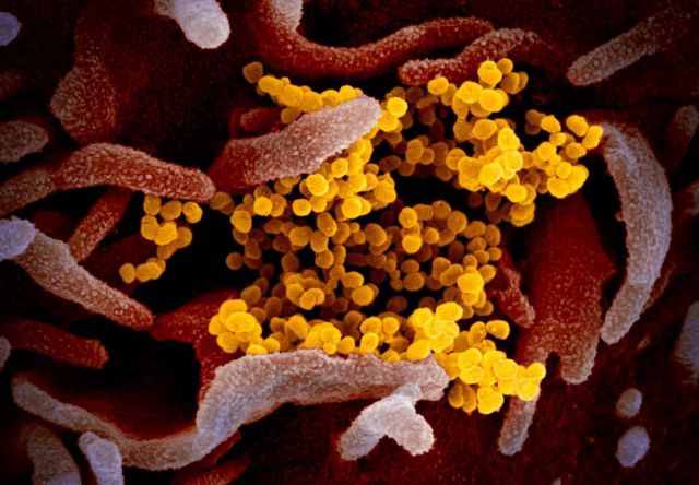
Scientists released stunning images of the Coronavirus, that they took with a variety of electron microscopes.
NIAID’s Rocky Mountain Laboratories (RML) in Hamilton, Montana, produced images of the novel coronavirus (SARS-CoV-2, previously known as 2019-nCoV) on its scanning and transmission electron microscopes on Tuesday, Feb. 11, 2020. SARS-CoV-2 causes COVID-19 disease, which has grown to be a global public health emergency since cases were first detected in Wuhan, China, in December 2019.
Above: This scanning electron microscope image shows SARS-CoV-2 (yellow)—also known as 2019-nCoV, the virus that causes COVID-19—isolated from a patient in the U.S., emerging from the surface of cells (pink) cultured in the lab. Credit: NIAID-RML
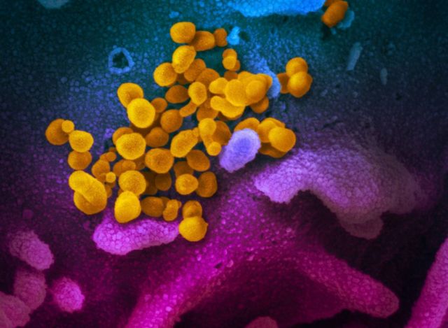 A scanning electron microscope image showing SARS-CoV-2 (yellow)—also known as 2019-nCoV, the virus that causes COVID-19—isolated from a patient in the U.S., emerging from the surface of cells (blue/pink) cultured in the lab. Credit: NIAID-RML
RML investigator Emmie de Wit, Ph.D., provided the virus samples as part of her studies, microscopist Elizabeth Fischer produced the images, and the RML visual medical arts office digitally colorized the images.
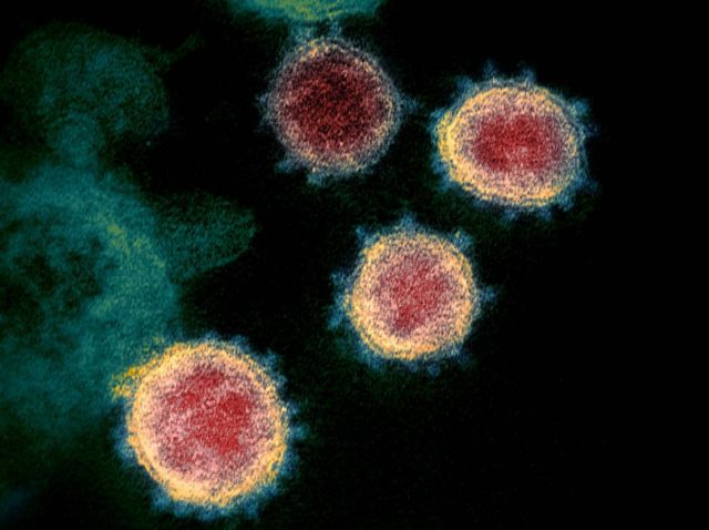 The spikes on the outer edge of the virus particles give coronaviruses their name, crown-like. Credit NIAID-RML
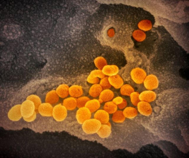 This scanning electron microscope image shows SARS-CoV-2 (orange)—also known as 2019-nCoV, the virus that causes COVID-19—isolated from a patient in the U.S., emerging from the surface of cells (gray) cultured in the lab. Credit: NIAID-RML
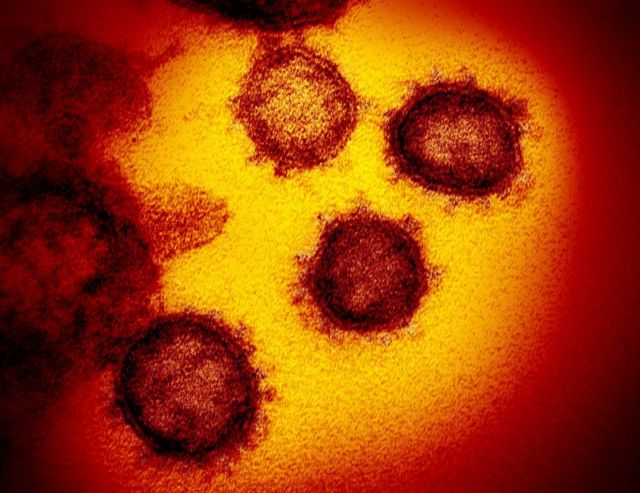 This transmission electron microscope image shows SARS-CoV-2—also known as 2019-nCoV, the virus that causes COVID-19. isolated from a patient in the U.S., emerging from the surface of cells cultured in the lab. Credit: NIAID-RML
Source: NIAID



Leave A Comment